Open or closed lateral canthoplasty often performed in conjunction with various facial rejuvenation procedures Taban OPRS 2010 eg upper- andor lower-lid blepharoplasty midface lift Contraindications. Mild lower-lid laxity or lateral canthal deformity.

Medial Canthal Webbing Seen After Upper Lid Blepharoplasy Done By A Download Scientific Diagram
Four procedures are described to repair these webs.

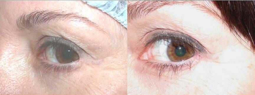
. The cosmetic result was highly satisfactory in all cases. And blepharoplasty represent the commonest iatrogenic causes of medial canthal webbing. Webbing 3 weeks after blepharoplasty.
At this point after surgery it is normal to have thickening and redness of the incision line. Seven 88 of the patients had lateral canthal webs after surgery and 1 12 patient had a medial canthal web after a motor vehicle accident. Webbing of the lateral canthus where outer upper and lower lids meet is called a canthal web.
Medial canthal webbing can be revised with a Z-plasty. 78 of the 171 patients with the inside out blepharoplasty had follow up of 3 months. Medial canthus Mohs defect.
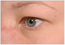
For an upper lid blepharoplasty ending the incision just lateral to the punctum avoids medial canthal webbing as well as lacrimal system injury. Large basal cell carcinoma. Download scientific diagram Medial canthal webbing seen after upper lid blepharoplasy done by a dermatologist.
In some cases early recognition and aggressive massage can result in better aesthetic results by improving. Although it appears that you have subtle webbing this appearance may be quite different after complete healing. Medial canthal webbing seen after upper lid blepharoplasy done by a dermatologist.
Aggressive excision of medial eyelid skin including some nasal skin spectacle deformation of the medial canthus and a preexisting tarsal fold increase the risk of web formation. If a line is drawn between medial and lateral canthi it will show an upward inclination to the lateral side. Post-blepahroplasty webbing can be seen when upper and lower blepharoplasty is performed together and the lateral skin incisions of the procedures are in close proximity less than 5 mm apart.
This area near the nose is called the medial canthus and the same area on the outer eyelids is called the lateral canthus. Intraoperatively the nasal end of the lid crease mark should not extend past the punctum or it should flare upward slightly after reaching the punctum. An effective preventive measure is to taper the nasal incision markings superiorly and to avoid extending the marking medially to the punctum.
Significant medial canthal tendon laxity see above. Postoperative canthal webbing is aesthetically displeasing and may be noticed by the patient andor surgeon. Ad When it comes to blepharoplasty or surgery of the eyelids there are certain things that.
Do it at least 3 times a day with 3 repeatitions and 20-30 seconds hold in each more Sorry about your problem. Prevent by planning an incision that extends to the medial commissure. A negative canthal tilt is present when the medial canthus is superior to the lateral canthus.
Medial canthus Mohs defect. During closure the running suture can be started just lateral to the medial-most aspect of the incision. Every patient voiced aesthetic concerns with the web and 4 57 of the 7 patients with lateral canthal webs.
Medial canthal webbing Attention to a few important surgical guidelines regarding medial upper eyelid wound closure will help prevent webbing in the medial canthus. Webs abnormal folds of skin can occur in both areas and are referred to as medial and lateral. Large basal cell carcinoma.
The best way to handle it is to do a push-pull massage of the surgical area. Canthal webbing can be associated with scleral show laterally due to inferior lid retraction and is a known complication of blepharoplasty or reconstruction following trauma or tumour excision 1. It provides excellent cosmesis and is associated with minimal complications.
I recommend Z-plasty repair of the medial canthal webbing. The experienced surgeon who is certain that the levator muscle and aponeurosis was identified and preserved during surgery will not be alarmed. Less Sorry about your problem.
Revisional Eyelid Surgery Fixing the Canthus. Canthal web revision Canthoplasty Revision Canthoplasty The area where the upper and lower lids meet is called the canthus. Secondary lateral canthal webs may occur after trauma or blepharoplasty.
May be due to inadvertent trauma to the levator complex including postsurgical edema and. This is called positive canthal tilt. May be due to incision extended too far medially.
The punctum is a useful landmark for the upper lid and lower lid incision. Incisions should be at least 45 mm above the punctum to avoid the canaliculus. After glabellar flap repair.
Normally the lateral canthus consists of an almond shaped angle. May be corrected by Zplasty Wplasty transposition flaps or YV advancement procedures. Two cases had minor webbing of the medial upper lid.
The rhomboid flap is an effective quick and simple technique for medial canthal reconstruction. WARNING Some photos may be explicit. Everyone has seen their eyes be tired and puffy but some people have to live with that.
There were no major complications or re-operations. After glabellar flap repair. Yes by an expert boarded PS.
Again the best treatment is prevention. In most patients the lateral canthal angle is positioned at 2 mm superior to the medial canthal angle. When webbing occurs skin obscures the angle and the canthal angle resembles a ducks foot which is normally webbed.
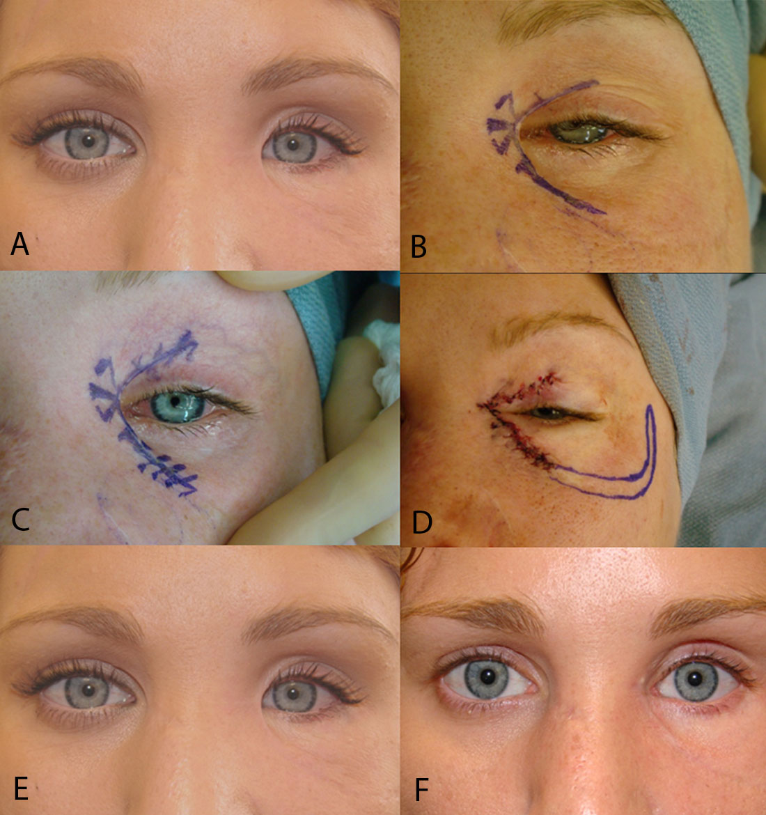
The del Campo technique 5. The scars usually occur when the incisions are carried too medially and the skin bridges the supero-medial hollow of the upper lid in a straight line. All clinical photographs are actual patients of Dr.
Determine the position of the lower eyelid and lateral canthus after release of the lower lid retractors with the inside-out technique by measuring the MR2 and use of the lateral canthal rounding scale. Ptosis of varying degree is common for patients to experience the day after upper lid blepharoplasty.

Medial Canthal Webbing Seen After Upper Lid Blepharoplasy Done By A Download Scientific Diagram
![]()
Medial Canthal Webbing Seen After Upper Lid Blepharoplasy Done By A Download Scientific Diagram
Case Of Bilateral Iatrogenic Medial Canthal Webbing Treated With Full Thickness Skin Grafts Medcrave Online
Case Of Bilateral Iatrogenic Medial Canthal Webbing Treated With Full Thickness Skin Grafts Medcrave Online
Revisional Eyelid Surgery Fixing The Canthus Dr Guy Massry
Medial Canthal Webbing Seen After Upper Lid Blepharoplasy Done By A Download Scientific Diagram

Medial Canthal Webbing Seen After Upper Lid Blepharoplasy Done By A Download Scientific Diagram
Canthal Web Revision Canthoplasty Revision Canthoplasty Dr Guy Massry
0 komentar
Posting Komentar